Routine tips
25 hospital microbiology procedures. Each one delivers the step-by-step technique and its clinical impact on the report.
Pre-analytical phase and Gram stain
No technique can save a poor-quality sample. This is where it is decided whether the result will mean anything to the clinician at the bedside.
- 01
Sputum: read the smear before plating
Bartlett score — more than 25 epithelial cells per low-power field indicates saliva, not lower-airway secretion. Reject the sample and request a new collection instead of processing it.
Why it matters: Avoids reporting oropharyngeal flora as a pneumonia pathogen and prevents unnecessary antibiotic use.
- 02
Urine outside the time window is a wasted sample
Up to 2 h at room temperature or 24 h at 2–8 °C. A container left at the nursing station for half a shift multiplies contaminants and destroys the specificity of the colony count.
Why it matters: The CFU/mL count is only meaningful if the sample was not incubating inside the container.
- 03
A dry swab is not an adequate specimen
Prefer aspirate or tissue fragment for wounds and abscesses. A superficial swab collects edge colonisers, not the pathogen driving the deep process.
Why it matters: It completely changes the interpretation of surgical-site and diabetic-foot wounds.
- 04
Gram stain is a quality screen, not just a morphology check
Record epithelial cells, polymorphonuclear cells and the predominant morphology. This combination already guides empirical therapy hours before culture results are available.
Why it matters: In sepsis and meningitis, the Gram stain is the fastest piece of information the laboratory can deliver.
- 05
Decolorisation: 3 to 5 seconds, timed
Excess alcohol-acetone turns a Gram-positive organism into a false Gram-negative. Always run a known control with the same stain lot.
Why it matters: A Gram-positive coccus misread as negative changes the entire empirical regimen.
- 06
Cross-check Gram stain and culture before releasing
A Gram stain showing cocci in clusters with a culture yielding only a Gram-negative bacillus is a red flag for a mixed-up sample, contamination or selective overgrowth. Investigate before signing off.
Why it matters: Unresolved discrepancies are the most common source of an incorrect report.
Blood culture
This is the test with the greatest clinical impact in the department, and the one most often undermined by collection details outside the laboratory's control — which the laboratory still needs to enforce.
- 01
Volume is the number-one factor for sensitivity
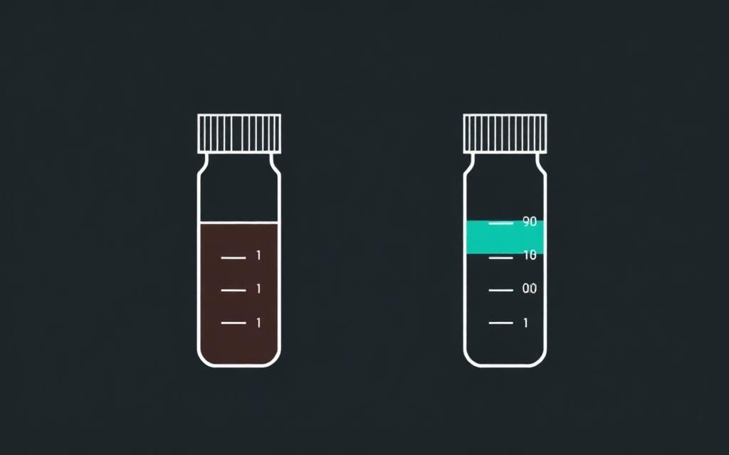
8–10 mL per bottle in adults. Each millilitre short reduces recovery by roughly 3%. In paediatrics, use the weight-based volume from the pediatric bottle protocol.
Why it matters: Adult bacteraemia is often below 10 CFU/mL — too little blood, no growth.
- 02
Two to three sets, different sites, before antibiotics
Separate venipunctures within the same time window. A single set does not allow contamination to be judged, and drawing after the first antibiotic dose greatly reduces positivity.
Why it matters: Without two sites there is no way to distinguish a contaminant from true bacteraemia.
- 03
Skin antisepsis with the correct drying time
Alcoholic chlorhexidine at 0.5–2%, friction and complete drying before puncture. Do not re-palpate the vein after antisepsis without a sterile glove.
Why it matters: Most coagulase-negative staphylococcus contamination originates in those 30 seconds.
- 04
Do not routinely draw from an indwelling catheter
Blood from a central line is used for the differential-time-to-positivity set when catheter-related infection is suspected, with a differential positivity time greater than 2 h — it does not replace peripheral venipuncture.
Why it matters: Catheters have lumen colonisation, which inflates false-positive results.
- 05
Time to positivity tells a story
S. aureus, Enterobacterales and yeasts usually turn positive within 24 h. A coagulase-negative staphylococcus that flags after 48 h, in a single bottle, looks like a contaminant.
Why it matters: It is a practical criterion to avoid treating a contaminant as a true infection.
- 06
A positive Gram in the bottle calls for a phone call, not just a report
Communicate the result immediately, recording the name of the physician on duty, the time and who received it. Log the communication in the system.
Why it matters: Advancing the regimen by hours changes mortality in sepsis — and a documented call protects the laboratory.
Urine culture and swabs
The highest-volume section, where interpretation — not technique — is the bottleneck. The question is never 'did it grow?', but 'does this growth mean infection?'.
- 01
Calibrated loop and the correct conversion factor
A 1 µL loop multiplies the count by 1,000; a 10 µL loop, by 100. Confirm which loop is in use before calculating — a silent loop swap is a classic on-call error.
Why it matters: A wrong factor shifts the result by one order of magnitude and crosses the breakpoint.
- 02
The cut-off depends on the collection method and the patient
Symptomatic midstream urine: ≥10⁵ CFU/mL, but ≥10³ already counts in a woman with cystitis and a single organism. Suprapubic aspirate: any growth counts. Catheter specimen: ≥10³.
Why it matters: Applying 10⁵ across the board causes the laboratory to miss real infections.
- 03
Three or more species = recollect
A polymicrobial culture from a midstream sample is perineal contamination until proven otherwise. Report it as such and request a new sample instead of identifying every organism.
Why it matters: Identifying a contaminant leads to untargeted antibiotic use and unnecessary cost.
- 04
Asymptomatic bacteriuria is not meant to be treated
Outside pregnancy and pre-urological-procedure screening, flag on the report that management depends on symptoms. Correlate with the urinalysis (leukocyturia, nitrite).
Why it matters: It reduces selective pressure and resistance across the whole hospital.
- 05
Tracheal aspirates need a quantitative count
Treat it quantitatively: ≥10⁵ CFU/mL for endotracheal aspirate, ≥10⁴ for bronchoalveolar lavage. Without a count, tube colonisation gets reported as 'pneumonia'.
Why it matters: This is what separates true ventilator-associated pneumonia from artificial-airway colonisation in the ICU.
- 06
Isolate before identifying
Streak until you obtain an isolated colony and subculture before any biochemical test or automated panel. A mixed colony invalidates both identification panel and antibiogram.
Why it matters: A panel run on a mixed culture generates a susceptibility profile that does not correspond to any real organism.
Antibiogram and resistance
The susceptibility report is the department's final product. Every technical detail here becomes a therapeutic decision within a few hours.

- 01
0.5 McFarland, inoculated within 15 minutes
A suspension left standing loses standardisation. Outside this window the inoculum changes and zone diameters shift in either direction.
Why it matters: A heavy inoculum produces false resistance; a light one produces false susceptibility.
- 02
Mueller-Hinton agar at 4 mm thickness
Thin agar enlarges zones, thick agar shrinks them. Use a dry plate with no excess surface moisture, and streak in three directions with a well-drained swab.
Why it matters: Half of all zone-diameter discrepancies come from the plate, not the strain.
- 03
Respect the distance between disks
No more than 12 disks on a 150 mm plate, 5 on a 90 mm plate, with 24 mm centre-to-centre spacing. Overlapping zones cannot be measured.
Why it matters: Overlap forces the test to be repeated and delays the report by 24 h.
- 04
Read the edge of complete inhibition, against the light
Ignore faint satellite colonies inside the zone — except for Proteus, where swarming should always be read as the outer edge. Measure from the back of the plate.
Why it matters: Reading a partial edge is the difference between susceptible and intermediate.
- 05
Confirmatory tests are not optional
D-test for inducible clindamycin resistance, double-disk approximation for ESBL, cefoxitin for MRSA, and carbapenemase screening (Blue-Carba/mCIM) whenever the profile calls for it.
Why it matters: Without a D-test, a 'susceptible' clindamycin result fails midway through treatment.
- 06
A critical profile means an immediate call to infection control
Carbapenemase producers, VRE, MRSA at a sterile site and unexpected multidrug resistance go out by phone and are logged in the same shift — they do not wait for the written report.
Why it matters: It triggers contact precautions before the patient's next contact with the ward.
- 07
Bank the strain and feed the local antibiogram
Store relevant isolates and periodically consolidate the institution's cumulative susceptibility report.
Why it matters: It is the local antibiogram — not the package insert — that defines empirical therapy in your hospital.
Standardise before trusting the result
Every procedure here assumes quality control of the media lot, current ATCC strains, and your service's SOP as the final word. If it diverges from the local protocol, the local protocol wins.